Adaptive optics microscopy for fine live imaging
1. Towards the non-invasive imaging of brain networks and functions at high resolution
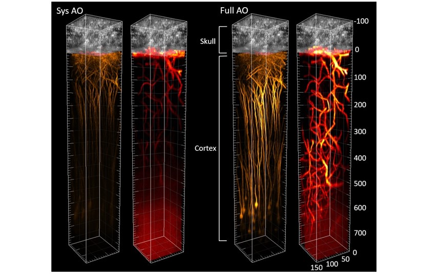
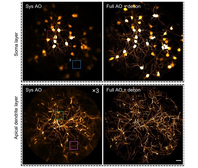
We developed a new microscope system combining three-photon excitation and adaptive optics capable of high-resolution in vivo imaging of fine neuronal structures in the mouse cortex through intact skull of mouse. We demonstrated the use of this platform to guide precise laser-mediated microsurgery through the intact skull and for accurate and sensitive functional calcium imaging of brain.
Related articles:
- Zhongya Qin, Zhentao SHE, Congping Chen, Wanjie Wu, Jackie K.Y. Lau, Nancy Y. Ip, Jianan Y. Qu (2022).
“Deep tissue multi-photon imaging using adaptive optics with direct focus sensing and shaping”. Nature Biotechnology, (2022)
- Congping Chen, Zhongya Qin, Sicong He, Shaojun Liu, Shun-Fat Lau, Wanjie Wu, Dan Zhu, Nancy Y. Ip, Jianan Y. Qu (2021).
“High-resolution two-photon transcranial imaging of brain using direct wavefront sensing”. Photonics Research, Vol. 9, Issue 6, pp. 1144-1156 (2021)
Related research grants:
- General Research Fund (16102123): “Rapid focus sensing and shaping for near non-invasive microscopy of the brain with high-resolution and a large field-of-view”
- General Research Fund (16102122): “Noninvasive deep brain imaging of adult zebrafish at high resolution”
- General Research Fund (16102518): “High-resolution imaging of mouse brain in vivo through thinned skull cranial window based on adaptive optics wavefront correction”
- Collaborative Research Fund (C6001-19EF): “High-resolution adaptive optics microscope system for live and deep imaging of biological tissues”
2. Deep brain imaging using adaptive optics two-photon endomicroscopy
Optical deep brain imaging in vivo at high resolution has remained a great challenge. We developed an adaptive optics two-photon endomicroscopy providing a minimally invasive approach to image buried brain structures at high-resolution. A new precompensation strategy plays a critical role to correct aberrations over large volumes and achieve rapid random-access multiplane imaging. We investigate the neuronal plasticity in the hippocampus, a critical deep brain structure, and reveal the relationship between the somatic and dendritic activity of pyramidal neurons.
Related article:
Related research grants:
- General Research Fund (16102518): “High-resolution imaging of mouse brain in vivo through thinned skull cranial window based on adaptive optics wavefront correction”
- Collaborative Research Fund (C6001-19EF): “High-resolution adaptive optics microscope system for live and deep imaging of biological tissues”
3. Adaptive optics two-photon microscopy for structural and functional retinal imaging
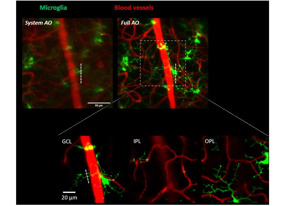
In vivo fundus imaging offers non-invasive access to neuron structures and biochemical processes in the retina. However, optical aberrations of the eye degrade the imaging resolution and prevent visualization of subcellular retinal structures. We developed an adaptive optics two-photon excitation fluorescence microscopy (AO-TPEFM) system to correct ocular aberrations based on a nonlinear fluorescent guide star and achieved subcellular resolution for in vivo fluorescence imaging of the mouse retina. The AOTPEFM permits structural and functional imaging of the mouse retina with submicron resolution.
Related article:
Related research grants:
- General Research Fund (16103215): “In Vivo Adaptive Optics Multiphoton Microscopy for the Study of Retinal Ganglion Cell Death and Glaucoma”
- General Research Fund (16102421): “In vivo visualization of structural and functional progression of retinal diseases at subcellular resolution in mouse models”